Menu

3D cell culture:overview and news

On this page, we give a brief overview over the basics of 3D cell culture with cell lines and primary cells. We describe differences between the classic 2D- / monolayer culture and 3D models. We give examples for 3D models, co-culture systems, matrices and fields of application in research, pharmaceutical labs and medicine.

The following topics are covered below:

3D Zellkultur MSC Differenzierung


3D cell culture basics

How do 2D and 3D cell culture differ?

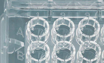
The normal standard type of cell culture is 2D- or monolayer culture of adherent cells in cell culture vessels like dishes, plates, multiwell plates, flasks etc. Hierdurch haben die Zellen in Kultur nur ein Oberfläche mit der sie Kontakt haben und die Nachbarzellen sind nur in 2 Ebenen (2D) neben den Zellen engeordnet. Diese Form nennt man auch Monolayer-Wachstum, da die Zellen in einer Lage auf der Oberfläche wachsen. In einem 3D-System haben die Zellen in allen 3 Ebenen Kontakte. Entweder nur zur Oberfläche oder zu Nachbarzellen rings herum.

3D systems are generated by culture in/on::

  • extracellular matrix (e.g. Matrigel)
  • gels of collagen, laktate or alginates
  • sponges of matrices
  • hanging drop method
  • in special culture vessels
    (e.g. transwells, spheroid plates)

Zellen in 2D und 3D Zellkultur

How do cells in 2D and 3D models differ?

2D and 3D culture models result in highly different cells and models. This means that neither the cells nor the results are same some times not even comparable. The following chracteristics and attributes of cells differ in the two model systems:

  • Cell morphology (structure)
  • Cell-matrix contacts (surface, mtarices)
  • Cell-cell contacts
  • Migration (potential, speed)
  • Proliferation capacity
  • Potential to differenciate (terminal differenciation and partial differentiation)
  • Development of cell polarity
  • Reaction to added compounds, drugs, test substances
  • Oxygen supply, pO2
  • Nutritional supply
Keratinocyten in 2D Zellkultur 3D Hautmodell


Advantages and disadvantages of 2D versus 3D cell culture?

In summary the following advantages and disadvantages of 2D/3D cell culture are known.

2D cell culture 3D cell culture
  • is fast
  • is much slower (some models take 6-8 weeks to grow)
  • produces high cell numbers
  • produces smaller cell numbers, models are more elaborate to grow
  • easy to use in high troughput
  • more difficult to use in high troughput, special material necessary
  • is cheap
  • is more expensive or very expensive
  • does not reflect the situation in tissues
  • reflects the tissue ebnvironment more closely
  • does not allow to study all processes
  • does allow to study more processes
  • cell lines are mostly used
  • most models need primary cells except in cancer reasearch


3D-cell culture models

Cells can be cultured in 3D in many differents ways or, models or settings. Below we summarized some of the most important culture systems and examples for 3D models:

  • Culture in transwells (only partially a 3D model)
    • Transport models with epithelial or endothelial cells
      • Polarization of cells (apical and basolateral polarization or luminals versus abluminal side)
      • Addition of compounds from both sides possible
      • Transport studies (across cell layers in blood vessels, blood brain barrier, lung epithelia etc.)
    • Migration models
      • Only one cell type
      • Migration (movement) of the cells through pores from one side to the other
      • Chemotaxis can be studied (e.g. Blood cells through blood vessel endothelia)
  • Co-culture systems in transwells (only partially a 3D model)
    • Blood brain barrier model
      • Usually two cell types in separate compartments (endothelial cells from brain microvessels and astrocytes)
      • Endothelial cells in the top compartment on a porous special membrane, astrocytes in the lower Transwell compartment
      • Compound addition from both sides possible, medium connests compartments
      • Transport studies across blood-brain barrier
      • Transmigration studies with additional blood cells
  • Sphäroids (cell spheres, solid or hollow)
    • Sphäroids in hanging drops
    • Sphäroids in gels (collagen, alginate, Matrigel®)
      • Evaluation of angiogenesis (formation of new blood vessels, atherosclerosis)
      • Inhibition of blodd vessel formation (cancer research)
  • Tissue engineering (Formation of tissues in culture or in vitro)
    • Skin models (epidermal or dermal, only keratinocytes or keratinocytes and fibroblasts)
      • Keratinocytes on collagen
      • Fibroblasts, collagen and kerationcytes
    • Skin transplants (micine, autologous=own skin is generated)
    • Vessel models with endothelial cells, muscle and fibroblasts)
    • Heart valve engineering (medicine, endothelial cells on matrix)
    • Bone models (medicine)
    • Cartilage models and transplants (medicine)
  • Organ models (Organoids)
    • Liver-organoids
      • Pharmaceutical research and drug testing as well as tissue transplant research
      • Studies of metabolic degradation of drugs
    • Artificial, autologous Liver (medicine, so far only experimental)

Sphäroid-Kultur


Special culture materials

In order to cultivate cells in 3D systems several special materials and substances are needed. This includes e.g.

  • Cell culture plastic
    • Transwells with different membranes and pore diameters
    • Slit-well plates for spheroid culture
    • Hanging-drops
    • Cell culture dishes with hydrophobic surfaces (spheroid formation)
  • Scaffolds, carrier materials
    • Collagen, alginate, lactic acid gels
    • Matrigel
  • Cell culture media
    • Special spheroid formation media
  • Bioreaktores
    • Mechanical stimulation of cultures needed for differentiation of cells
      • Heart valve bioreactors
      • Cartilage bioreactors

PHCbi 3D cell culture 96 slit well plate


Examples

Generally spoken, sphäroids may be generated by 3 diffenrent mechanisms. Either they are plated on hydrophobic plastic surfaces that do not allow for adhesion, or cells are plated in hanging drops which do not offer any surface besides the membrane of the other cells because the cells sink into the hanging drop of medium, or öastly cells are cultured in special media that induce spheroid formation even on normal cell culture plastic surfaces that would allow for adhesion. One example, special hydrophobic plastic from PHCbi is shown below.

Sphäroid-Herstellung


We use cookies on our website. Some of them are essential for the operation of the site, while others help us to improve this site and the user experience (tracking cookies). You can decide for yourself whether you want to allow cookies or not. Please note that if you reject them, you may not be able to use all the functionalities of the site.